Challenges with interpreting ECG scan with LLM models

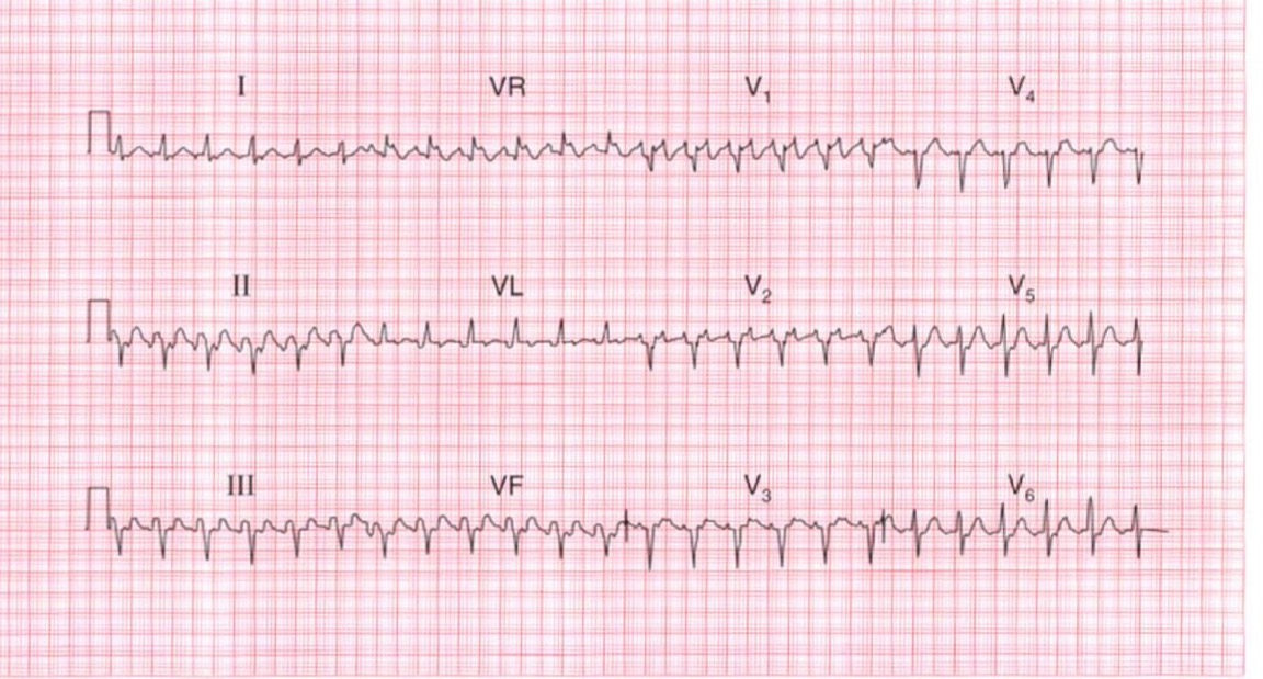
Large Language Models (LLMs) face several unique challenges when interpreting ECG scans, particularly in distinguishing between similar arrhythmias like Atrial Fibrillation and SVT. These challenges include:

  • Pattern Recognition Limitations: While LLMs excel at processing textual descriptions, they may struggle with subtle visual patterns in ECG waveforms that human cardiologists can readily identify.
  • Context Integration: Different models may weigh various ECG features differently, leading to varying interpretations. For example:
    • Some models might prioritize R-R interval regularity
    • Others might focus more on baseline characteristics or P wave morphology
    • The presence of noise or artifacts can affect different models in varying ways
  • Training Data Bias: Models may be trained on datasets with varying proportions of A-fib and SVT cases, leading to potential bias in their interpretations.
  • Feature Extraction Challenges: In cases where the distinguishing features between A-fib and SVT are subtle (like slightly irregular vs regular rhythms), different models may have varying thresholds for what constitutes "irregular" rhythm.

This case study demonstrates these challenges by examining how different interpretations can arise from the same ECG, particularly in distinguishing between Atrial Fibrillation with RVR and SVT, where the key differentiating features can be subtle and open to varying interpretations.

Atrial Fibrillation

Here are the most important points about the differential diagnosis between Atrial Fibrillation (A-Fib) with Rapid Ventricular Response (RVR) and Supraventricular Tachycardia (SVT) in this ECG. Let's compare them:

Similarities:

  • Rapid Rate: Both A-Fib with RVR and SVT present with a rapid heart rate.
  • Narrow QRS Complexes: Both typically have narrow QRS complexes, indicating a supraventricular origin.
  • Difficulty Identifying P Waves: In both, clear P waves can be difficult to identify due to the rapid rate.

Differences:

  • Rhythm:
    • A-Fib: Characteristically, A-Fib has an irregular rhythm.
    • SVT: SVT typically has a regular rhythm.
  • Atrial Activity:
    • A-Fib: In A-Fib, you often see fibrillatory waves, which are irregular, rapid oscillations of the baseline.
    • SVT: In SVT, there may be retrograde P waves (P waves that occur after the QRS complex), or the P waves may be buried within the QRS complexes, making them hard to see. If there is clearly visible atrial activity, it will tend to be much more regular than in A-fib.
  • ECG presentation of the Baseline:
    • in A-fib there will usually be visible fibrillatory baseline.
    • in SVT the baseline tends to be much more isoelectric.

Why the Distinction Matters:

  • Treatment: The treatment for A-Fib with RVR and SVT can differ.
  • Underlying Causes: The underlying causes can also be different.

In the context of the analyzed ECG:

My initial interpretation pointed towards A-fib because of the irregular R-R intervals, and the visible fibrillatory base line.

The interpretation, "Supraventricular Tachycardia (SVT). The rapid oscillations between the QRS complexes are suggestive of atrial activity, but the absence of clear P waves is characteristic of SVT." Is a valid interpretation. If there was a higher level of regularity to the R-R intervals, the SVT diagnoses would have been much more likely.

Conclusion:

Both A-Fib with RVR and SVT can present with rapid, narrow-complex tachycardias. The key differentiating factor is usually the regularity of the R-R intervals and the morphology of any visible atrial activity. Therefore, while both interpretations are valid, given the irregularity of the prior ECG, A-fib is the most likely.

Note: This is a demo version showcasing the core functionalities of the ECG Analyzer. It is not a substitute for professional medical evaluation and should be used as an aid to support clinical decisions.