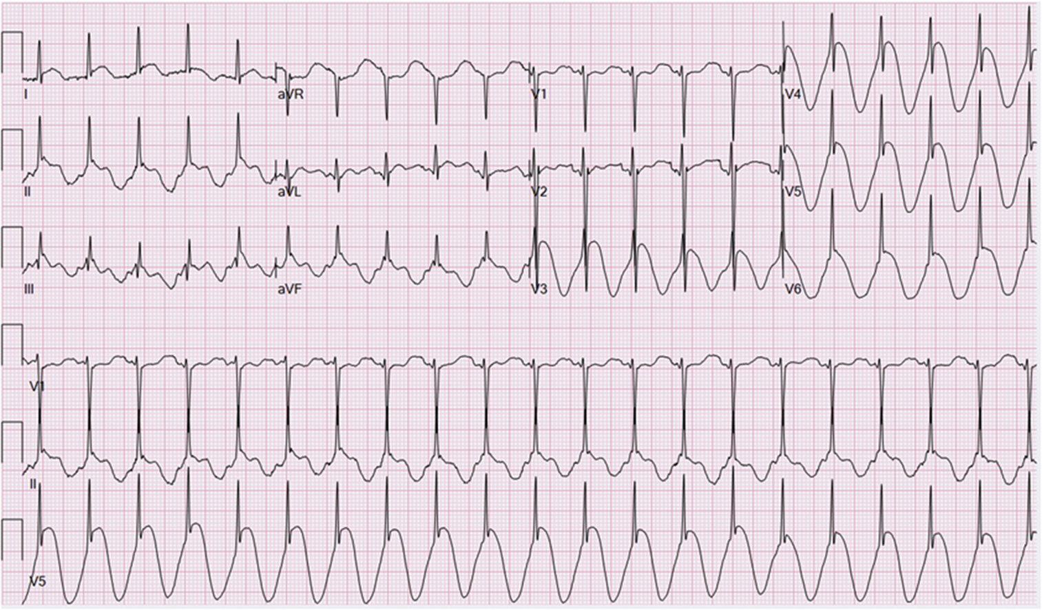
Description: Possible Left Bundle Branch Block (LBBB). The ECG shows a regular rhythm with a heart rate around 60 bpm and significantly widened QRS complexes.
Case Studies
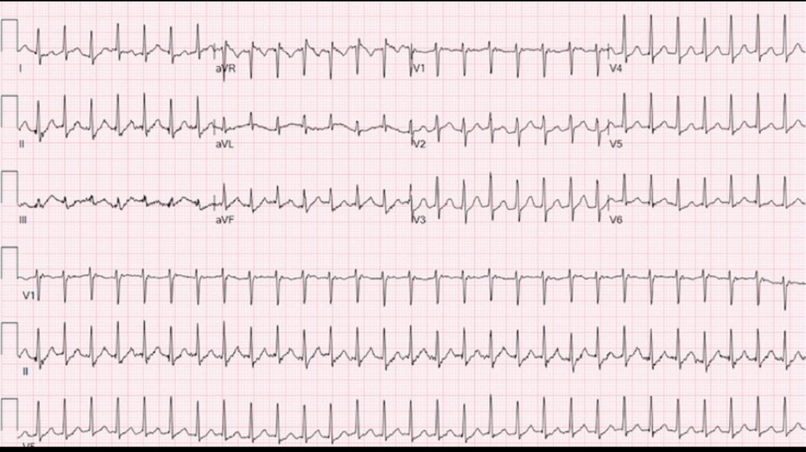
Description: Possible sinus tachycardia. The ECG shows sinus tachycardia with a heart rate of 125 bpm.
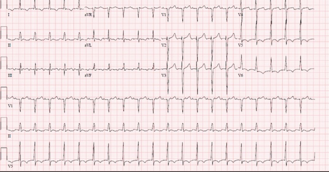
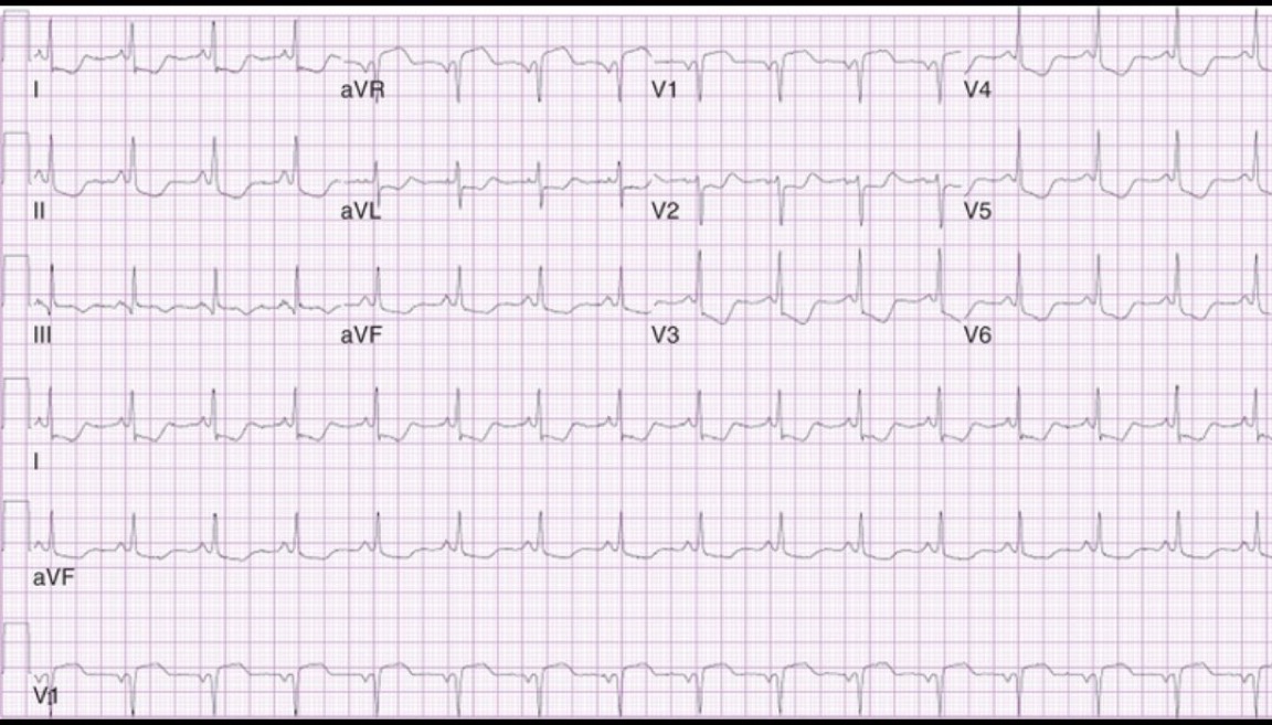
Description: The ECG demonstrates atrial tachycardia with a 2:1 atrioventricular (AV) block. The P waves in lead III, which are different from sinus P waves, suggest an ectopic atrial focus. The 2:1 AV block is most clearly seen in lead V1.
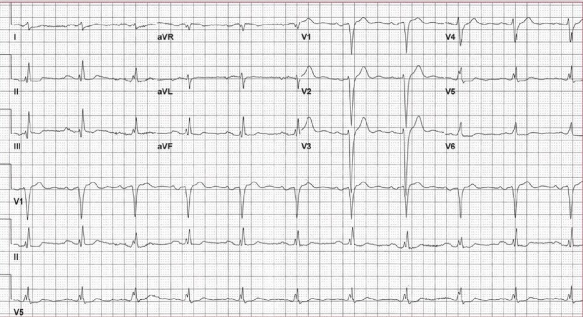
Description: Heart Rate: Approximately 50
bpm, indicating bradycardia.
Rhythm: Regular sinus rhythm.
Overall Interpretation: Sinus bradycardia with possible ST
segment depression requiring further evaluation.
Description: The ECG indicates a normal sinus rhythm with no significant abnormalities. However, to provide a comprehensive interpretation, it is essential to compare the findings with the patient's age, symptoms, and medical history. This ensures that subtle variations in the ECG are not overlooked and that any potential risks or conditions are appropriately evaluated.
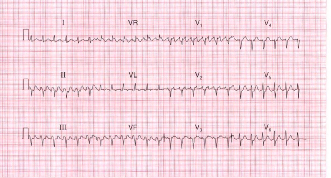
Description:This ECG demonstrates a very fast, irregularly irregular, narrow complex tachycardia. The absence of discernible P waves and the presence of a chaotic baseline strongly suggest Atrial Fibrillation with a Rapid Ventricular Response (RVR)